This post is also available in:
![]() العربية
العربية ![]() हिन्दी
हिन्दी
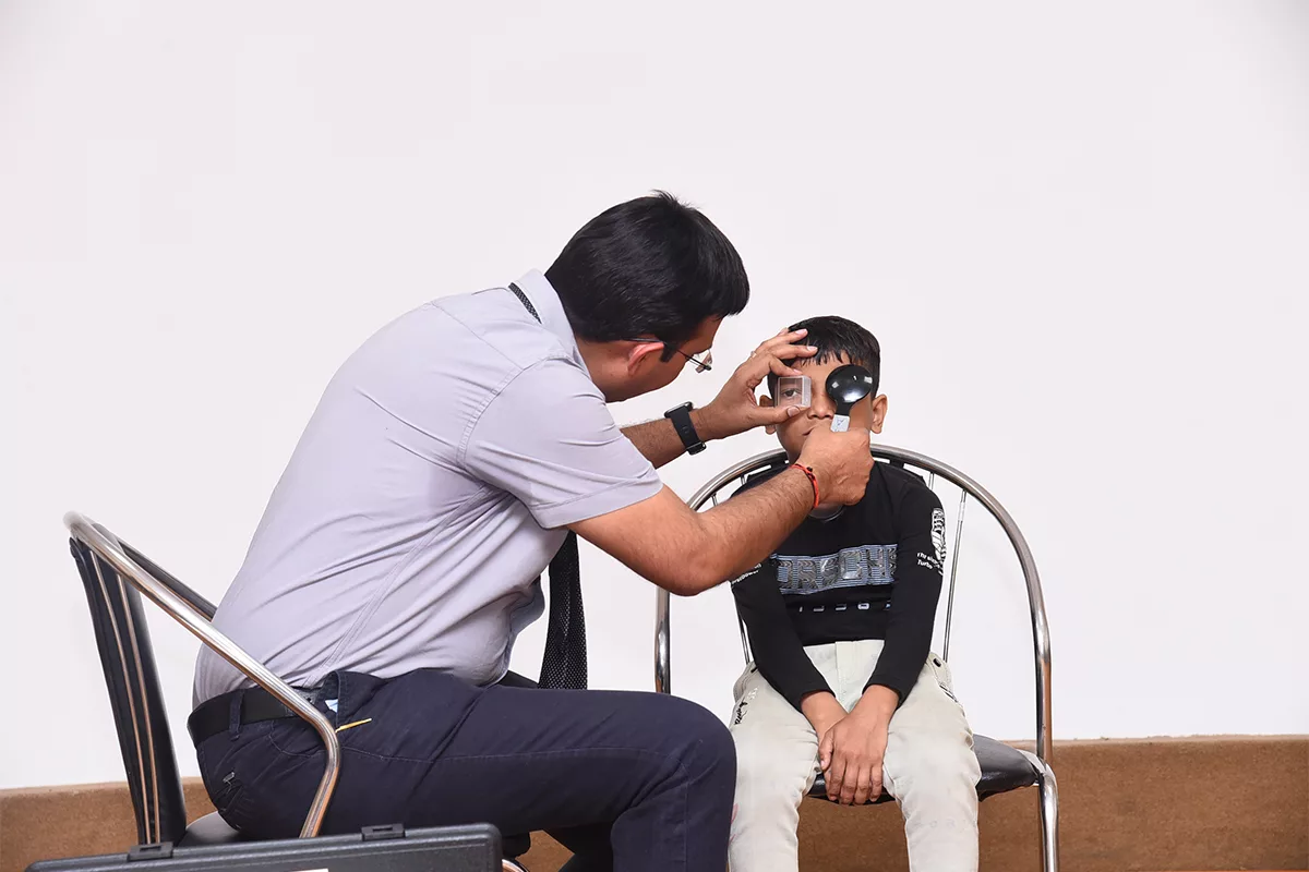
Take hints from what your eyes say, before it’s too late. We have Dr. Anasua Ganguly Kapoor. Consultant Ophthalmic Plastic Surgery and Ocular Oncology, LV Prasad Eye Institute, Vijayawada, India telling us what our eye symptoms may mean…
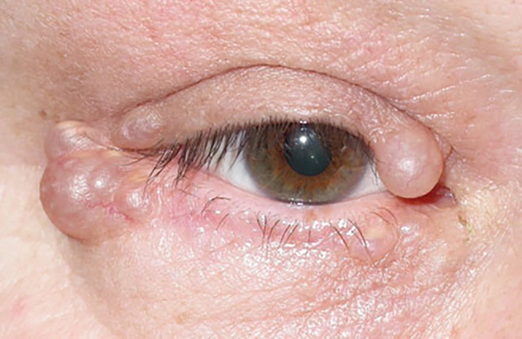
1.Lid nodule – Eyelid is the layer of skin which covers and protects the underlying eyeball. A lump or nodule on the eyelid is most commonly a sign of either inflammation/infection or rarely a tumor which can be benign (non-cancer) or malignant (cancer).
Causes of eyelid nodule:
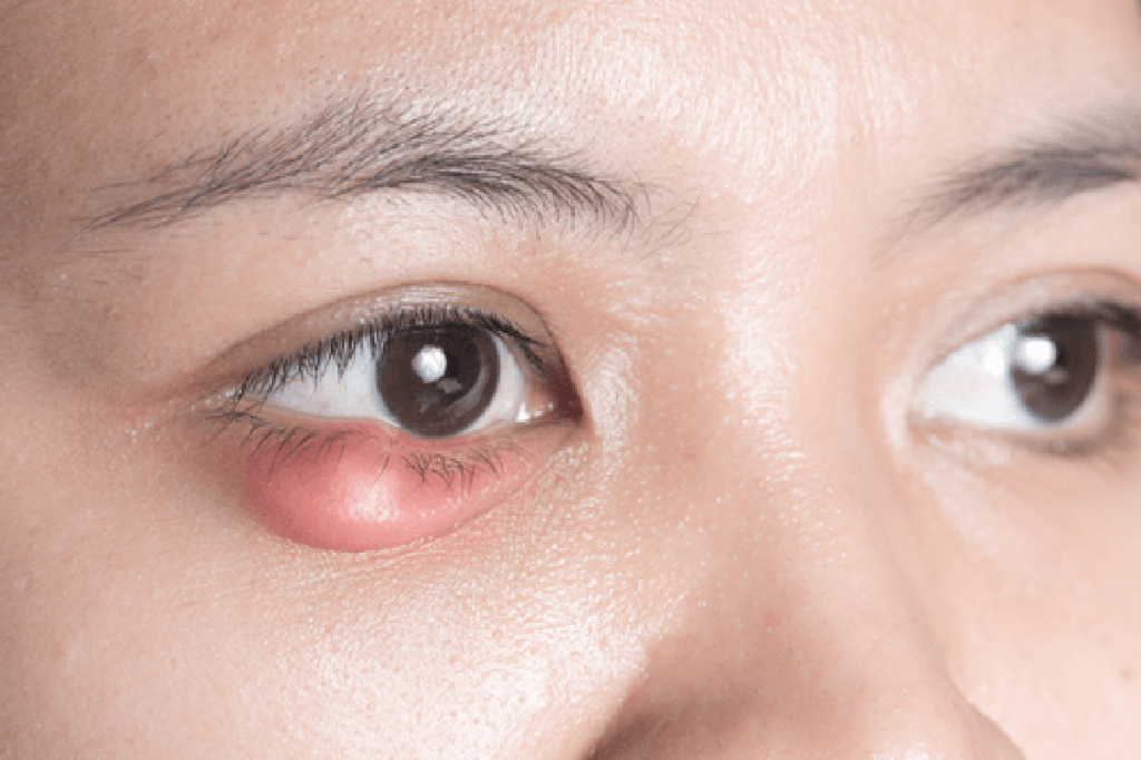
Stye is an acute painful red nodule on the eyelid margin, sometimes with a yellow spot indicating pus-pointing. The pain is usually out of proportion to the appearance. It develops because of bacterial (Staphylococcus) infection of the eyelash hair follicle gland (glands of Zeis).
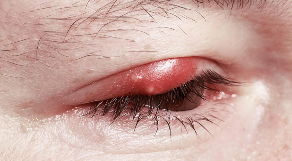
Chalazion: is a painless hard round lump on the eyelid usually a little away from the eyelid margin. It is typically painless and usually present for weeks or months before the patient seeks medical help. It is caused because of blockage of the oil glands of the eyelid (meibomian glands) and accumulation of secretions within. Frequently the patients have a previous history of similar lesions as they tend to recur in predisposed individuals.
The different possible predisposing conditions leading to these nodule formations are- Working in dusty environments, uncorrected eye refractive errors, excessive near work, acne rosacea, spread from local infection like blepharitis, conjunctivitis, seborrhea and systemic diseases like malnutrition and diabetes mellitus.
The different benign eyelid tumors can present as a water bubble like structure on the eyelid (cyst), pigmented finger like projections with surface irregularities (warts, keratin horns), a small pigmented elevation on the lid margin (eyelid nevus) or yellowish flat disc-like bumps around the eyes (Xanthelesma). However, it is important to differentiate these harmless eyelid bumps from malignant eyelid cancers. The danger signs are recurrent swelling at the same site, loss of eyelashes, surface ulceration or bleeding on the lump, thick blood vessels on surface or involvement of eyelid both inside and outside.
Treatment:
Stye and chalazion are usually harmless and heals on its own within a week, but it is safe to consult an eye doctor for proper diagnosis and early healing. You can encourage faster resolution by applying warm compresses for around 10 minutes 3 to 4 times a day. Antibiotic eye ointment application on the eyelid margin may be added for faster recovery. In case of persistent chalazion intralump injection of steroids or giving a small nick on the inner surface of the lesion may be considered.
The benign eyelid lesions may be observed as harmless. However, if you have cosmetic concerns or the lump blocks your vision, it can easily be removed. For malignant lid lesions, it is important to get it treated by an eye cancer specialist with complete removal of the lesion with some amount of normal skin margins followed by plastic surgery of the eyelid.
2.Eyelid Twitching – The muscles around the eye are usually under voluntary control. Eye or more accurately eyelid twitching is a condition where these muscles function abnormally and are not under direct control by the brain.
Types of eyelid twitching:
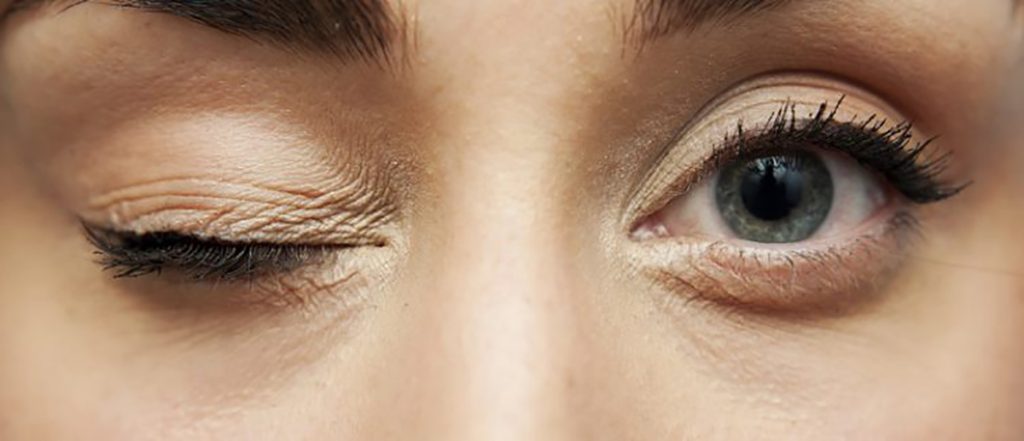
Benign eyelid twitch (Myokymia)– These are fine fluttering movement generally affecting one eyelid usually the lower eyelids or at times the upper eyelid. Affected person may feel a jumping movement on the eyelid whereas observers may barely notice the movement. This twitching is usually episodic lasting for seconds to hours or at times for months but eventually resolves on its own without any treatment. It may be associated with fatigue, stress, alcohol and caffeine.
Benign essential blepharospasm (BEB) – In this condition both eyes forcefully close at the same time intermittently. It may be associated with difficulty in opening the closed eyelid voluntarily (apraxia of eyelid opening). It occurs due to uncontrolled nerve signals in the brain stimulating the eye muscles. The spasms usually decrease during sleep and may be variable in severity from moment to moment and with different physical activity. Features of BEB may be associated with simultaneous writhing movements of the cheek, mouth, tongue or neck (Meige syndrome).
Hemifacial spasm – In this condition there is intermittent frequent contraction of muscles of one side of the face. It is caused by irritation of the facial nerve on one side leading to the spasms.
Secondary blepharospasm– Sometimes the forced eyelid closure is because of eye irritation (most commonly dry eye) or some medications (psychiatric drugs).
Treatment:
Persistent twitches may require treatment as frequent closure of the eyelid causes visual interruption in patients affecting their day to day activities. There is no cure for blepharospasms and treatment is only symptomatic. Neurologists may prescribe oral medications with limited help to some patients. Botulinum neurotoxin injection is most effective treatment available at present for temporary reduction of symptoms. Injections are administered into the affected areas as an outpatient procedure. However, patients not responsive to oral medications or botulinum are candidates for myectomy surgery where removal of muscles responsible for eyelid closure is performed by ophthalmic plastic surgeons. The last option in severe resistant cases is neurosurgery.
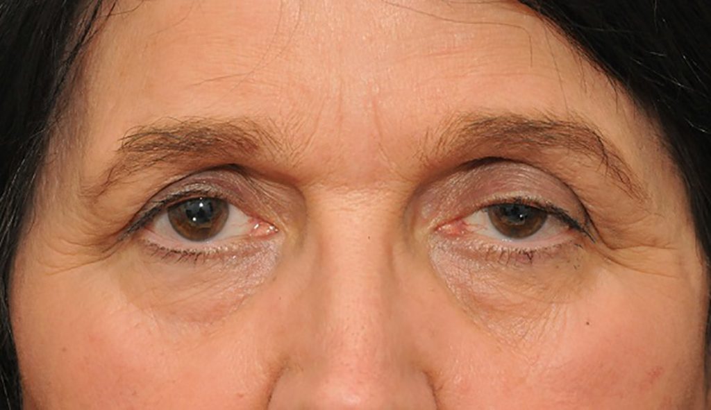
3.Droopy Eyelids– Normally our upper eyelid covers around 2mm of the cornea (the circular transparent layer in front of the eye). Whenever the upper eyelid comes below this level the condition is known as droopy eyelid or ptosis.
Causes of droopy eyelids:
Droopy eyelid can occur as a result of birth defect, muscle or nerve disorder, injury, lump pressing on the eyelid or simply due to aging.
Problems caused because of droopy eyelids:
Droopy eyelids give a sleepy, tired and aged appearance, and can even obstruct vision if severe. It may also cause excessive forehead wrinkling, elevated eyebrow, headache, eye fatigue, loss of peripheral visual fields, abnormal head posture such as chin elevation. In children severe droop might obstruct development of vision leading to a lazy (amblyopic) eye thus reducing vision. In adults, droopy eyelid is mainly a cosmetic concern.
Treatment:
It is extremely important to establish the cause of ptosis by an ophthalmic plastic surgeon to decide on the treatment. If there is an underlying cause it needs to be treated first. However, treatment for most cases of ptosis is surgical. Glasses that can hold the eyelid up (crutch glasses) are another option for patients who are not good candidates for surgery.
Timing of surgery:
In children surgery is often delayed till 4-5 years of age when child is cooperative for accurate preoperative ptosis measurement. Visual monitoring with an eye doctor is of utmost importance while the child is waiting for surgery at an interval of 3-12 months depending on the severity of ptosis to exclude the possibility of development of lazy eye. However, where ptosis covers the visual axis, surgery is advised early to prevent development of lazy eye and subsequent reduction of vision. Adults can undertake the surgery whenever they develop significant cosmetic concerns.
Surgical techniques:
Several surgical options are available for ptosis, and the type of surgery depends upon the severity of the droop. A simple lift of the tissues is performed from the back of the eyelid (Mullerectomy) in cases with mild eyelid droop. This is a scar less surgery. Moderate droop requires tightening of the muscle that lifts the eyelid (levator) and is preferably done from the skin side. This scar remains hidden in the upper eyelid skin fold. In severe droop, tightening the eyelid muscle may not be effective, and hence the eyelid must be connected to the forehead muscle (frontalis) with a string material to ensure eyelid lift. This surgery (called tarso-frontal sling surgery) involves small scars over the eyelid, just above the brow and forehead. Sling surgeries are not permanent, and the eyelid often droops in a few years due to blinking and gravity necessitating possible revision surgeries over his/her lifetime. Often, ptosis surgery needs one or two sessions of suture adjustment (1-6 weeks after surgery) to match it to the other eye. In case of severe ptosis, correction is optimal only in straight gaze. The eye appears slightly wider in downgaze. During sleep closure may also be incomplete.
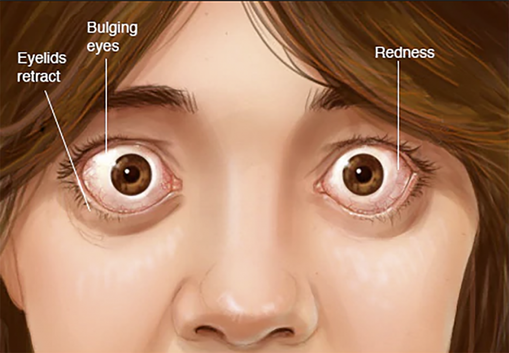
4.Bulging Eyes – Normally our upper eyelid covers around 2mm of the cornea (the circular transparent layer in front of the eye) and lower eyelid just touches the cornea. So bulging eyes are identified by visible white of the eyes above or below the cornea. Some people have big eyes since birth which is normal for them. So big eyes should not be confused with abnormally bulging eyes.
Causes of Bulging Eyes :
Bulging eyes can be a sign of an underlying serious medical disorder and mandates visit to an eye doctor. Eyes protrude from the eye socket either because of swelling of the muscles, fats or tissues behind the eye because of thyroid disorders (most common cause) or infection/ inflammation, or because of a lump (cancerous/ non-cancerous) developing in region behind the eye pushing the eye forward. Rarely injury to the eye may cause blood to collect behind the eye causing bulging.
Problems:
Bulging eyes can expose the cornea to dryness, making it prone for serious infection which may lead to reduction of vision. In severe cases it can even put the optic nerve under pressure leading to vision loss. Apart from these sight threatening problems there can also be associated double vision, pain, redness, swelling, dry eyes, discomfort, foreign body sensation.
Treatment:
Treatment of bulging eyes depends on the problem triggering it. Overactive thyroid gland because of an autoimmune disease (Graves’ disease) is the most common cause of bulging eyes. This disease usually has two stages and the treatment depends on the stage of the disease. In the initial active stage (first 18-24 months) treatment is with medications (lubricant eyedrops, oral and intravenous steroids if necessary). However, the bulging eyes usually persist in the inactive phase and requires surgery to expand the orbit (orbital decompression) for the eyes to fall back. In cases of infection or inflammation treatment involves control with oral or intravenous medications. In cases where the bulging is caused because of a lump behind the eye treatment involves surgical removal of the lump (orbitotomy) and confirmation of the nature of the lump by pathology examination. Further treatment in these cases depends on the nature of the lump on biopsy.