Five Essential Corneal Tests for Accurate Screening of Refractive Surgery Candidates
Accurate corneal evaluation to determine candidacy and establish what procedure is best for each patient is as important as the procedure itself.
We have come a long way from topography and ultrasound pachymetry of 30 years ago. As the field of refractive surgery matured, new methods of examining cornea have been added. We now have five different methods to evaluate cornea prior to refractive surgery. Each of these methods may have false positives and false negatives. Each is prone to artifact, from subtle angle kappa, to tear film, to human error in performing each test. It is, therefore, essential that we don’t just rely on several of these tests, but perform all five for each patient to accurately distinguish between healthy and structurally robust cornea vs. pre-ectatic vs. subclinical epithelial basement membrane dystrophy vs. contact lens warpage. Correct diagnosis is essential to successful outcome and great vision long term.
At Pacific Vision Institute, we see patients who have gone to multiple consults and received conflicting opinions. We often find that performing all five corneal tests invariably establishes a clear, definitive diagnosis and treatment plan for each patient.
Topography is usually the first corneal test performed. In this test, we look for inferior steepening, skewed axis and other possible irregularities.
Tomography is performed to evaluate anterior corneal surface further, to determine corneal thickness, including the exact location of the thinnest spot of the cornea, to evaluate posterior cornea, and to perform ectasia risk assessment with advanced software that compares multiple parameters of the patient’s cornea to those found in a general population of people with normal corneas.
Epithelial Thickness Mapping (ETM) is performed to evaluate a patient’s corneal epithelium to rule out subtle pre-ectatic conditions, contact lens warpage, and epithelial basement dystrophy. It is not uncommon for subtle focal abnormalities on topography and tomography to be misdiagnosed as pre-ectatic conditions, whereas they are truly epithelial basement membrane or contact lens warpage (Schallhorn, JM, et al. Distinguishing between contact lens warpage and ectasia: Usefulness of optical coherence tomography epithelial thickness mapping. J Cataract Refract Surg. 2017;43(1):60-66.) Topography and tomography simply do not show the “whole picture” and often lead the examiner to recommend PRK for patients who are great LASIK candidates based on ETM or recommend no procedure to patients who excellent PRK candidates. I have seen patients for second and third opinion whose diagnosis and treatment plan becomes unequivocally clear after performing and analyzing their ETM.
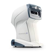 Ocular Response Analyzer (ORA) is a sophisticated technology designed to test the elasticity of the cornea and its ability to remain strong and stable after Laser Vision Correction. ORA is also helpful in sorting out different opinions about a patient’s diagnosis and treatment options and resolving split opinions. Moreover, a recent review of literature by Moshirfar M, et al has concluded that technologies such as ORA are essential for screening of corneal ectasia because “changes in biomechanical properties may occur before disease becomes apparent via tomography or topography.” (Moshirfar M, et al. Advances in biomechanical parameters for screening of refractive surgery candidates: A review of the literature, Part III. Med Hypothesis Discov Innov Ophthalmol. 2019;8:219-240).
CONTOURA® Vision Analysis: is performed to generate a highly detailed map of corneal higher order aberrations by measuring elevation profile of 22,000 unique data points on the cornea. This creates a highly detailed contour scan of microscopic peaks and valleys to screen out pre-ectatic conditions and to determine if the patient has been out of contacts for a sufficient length of time to establish a precise treatment plan.